Second Opinion on an MRI: You Already Have the Images
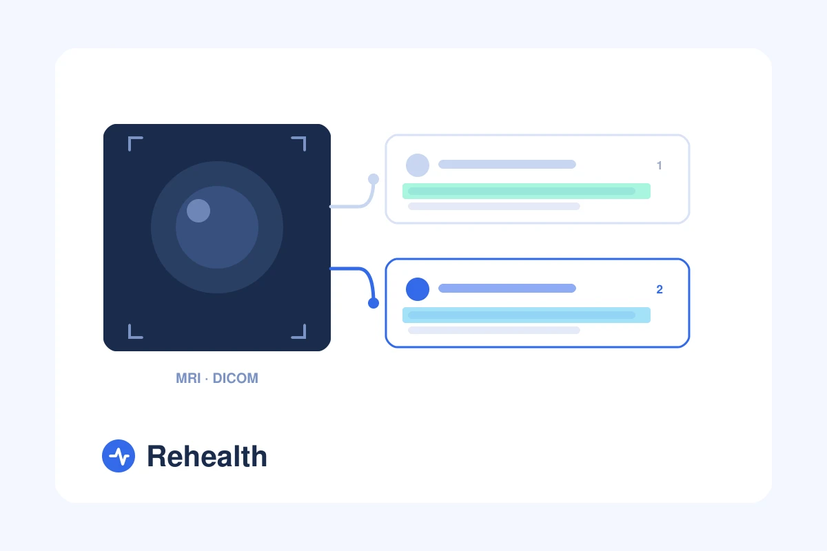
A second opinion on imaging does not mean new scans. The pictures you already have can be read a second time, by a second specialist.
You already have the images. What is missing is a second reading.
You had the MRI. You have the report. The file is sitting on a disc, in a portal, or in an email from the hospital.
Many people think a second opinion means starting again. New scans, new appointments, new bills. For imaging it usually does not. The pictures you already have can be read a second time.
In short
| 7.7% | of brain and spine scans re-read at one hospital got a different reading [1] |
| 26.2% | of bone and joint scans re-read at another had a clinically important difference [2] |
| 0 | new scans you need for a second reading |
A scan does not diagnose. A doctor reads it.
This is the part people miss. The machine makes pictures. It does not decide what they mean.
A radiologist looks at those pictures and writes a report. That report is one specialist's reading of what is there. Another specialist can look at the same pictures and describe them differently.
So the report in your hand is not a measurement. It is an opinion, written by a doctor, about images that stay the same.
How often does a second reading change something?
Two studies give a useful sense of the range.
At Johns Hopkins, 4,534 brain and spine scans sent in from other hospitals were read again. 347 of them, or 7.7%, got a different reading [1].
A second study looked at 2,326 bone and joint scans re-read by specialist radiologists. 610 of them, 26.2%, showed a difference that mattered for treatment. In cancer cases the figure was higher, 36.3% [2].
Read these numbers carefully. Both studies looked at scans sent to a specialist centre for review. The cases were often complex ones. They are not the error rate of your local hospital. What they do show is that a second reading is not an empty exercise.
Asking for a second reading is not a complaint about your doctor
This stops a lot of people, and it should not.
Radiology is a reading job. Two trained specialists can look at one knee and describe it in two ways. That happens in medicine every day. Both readings can be careful and still differ.
Often the second reading agrees with the first. That is a useful answer too. You stop wondering whether something was missed.
You do not need new tests
For a second reading you need two things, and you probably have both.
- The images themselves, not only the written report. These come as DICOM files, on a disc or through the hospital portal.
- The radiology report, plus the referral letter if you have one.
Blood tests and previous scans help, because a change over time often matters more than a single picture. But you do not book a new MRI. That saves the scan cost, the waiting time, and another dose of contrast.
When a second reading is worth it
Not every scan needs one. These are the moments when people ask:
- A cancer diagnosis or staging, where the plan depends on what the images show. See oncology and cancer care.
- Before joint surgery, when you are not sure the operation is the only option. See orthopaedics and joint surgery.
- An incidental finding, something the scan showed that nobody was looking for.
- Two reports that do not say the same thing.
- A report you were given but never had explained.
The pattern is the same in all of them. A decision is coming, and it rests on how the images were read.
How to get your images
Hospitals hold the images, but the file is yours.
In Europe you have the right to a copy of your own medical records. Ask the medical records desk or the radiology department. Say clearly that you want the images, not only the report. Most hospitals hand them over on a disc or through the patient portal, usually within a few days.
If the portal only shows the report, ask again for the DICOM files. The wording matters.
The scan is done. The reading can be done again.
Upload the images you already have and speak to a specialist online. From €35.
Get a second opinionHow it works at Rehealth
You upload the file in the app, not by email. It is handled under European data rules, and you choose what to share.
You pick the doctor yourself from the list of doctors. Every doctor sets their own fee, and prices start at €35.
The doctor reads your images and your report before the call. So the time is for your questions. Your own doctor at home keeps your case, and you can take the answers back to them. If treatment abroad comes up later, you can read how treatment abroad works.
Frequently asked questions
Do I need a new MRI for a second opinion?
No. A second reading uses the images you already have. A new scan is only needed if the doctor says the existing one is too old. The same goes for poor image quality.
Is the report enough, or do you need the images?
The images are the important part. The report is one doctor's reading of them. Without the images, a second doctor can only comment on someone else's words.
My hospital gave me the report but not the images. What can I do?
Ask the radiology department or the medical records desk for the DICOM files. In Europe you have the right to a copy of your own records. Most hospitals provide them on a disc or through the patient portal.
Does a second reading mean my radiologist made a mistake?
No. Reading a scan is interpretation, and two specialists can describe the same images differently. A second reading often confirms the first.
How old can a scan be?
It depends on the problem and on what changed since. Bring the dates of your scans to the call. The doctor will say whether the images still answer the question.
What do I get at the end of the call?
You receive clear next steps: what the images show, what the options are, and whether anything else needs looking at.
References
[1] Zan E, Yousem DM, Carone M, Lewin JS. Second-opinion consultations in neuroradiology. Radiology, 2010;255(1):135-141. View the study →
[2] Chalian M, Del Grande F, Thakkar RS, Jalali SF, Chhabra A, Carrino JA. Second-opinion subspecialty consultations in musculoskeletal radiology. American Journal of Roentgenology, 2016;206(6):1217-1221. View the study →